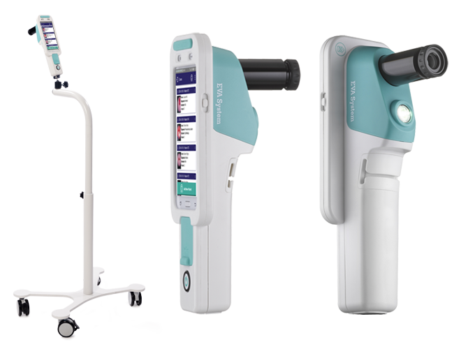
Description
Powerful Platform
A powerful hard core is a promise of excellent image performance.
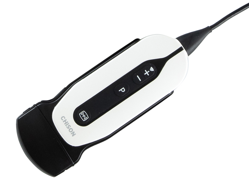
Ultrasound Platform — Holo
The Holo Platform provides an advanced 64-ray image. Due to the high-speed hardware platform, the system can handle up to 5000 frames/s, which means around 1GB/s of data is processed instantly. All of them dramatically increase diagnostic confidence and improve accuracy.
Hello PW
3 real-time image sampling gates. Allow the user to move each gate pre and post-processing. An essential tool for accurately assessing plaque. Used before, in and after the vascular position of pathological change with a synchronized measurement of a heartbeat.
versatile application
Advanced imaging technologies guarantee every quality exam.
Auto Doppler
The Wisonic Clover has the patented ability to automatically locate blood vessels. Clover automatically adjusts the color box position, offset angle, PW gate size position, PW offset angle, and correction angle. With Auto Doppler, confidence and time savings are a standard feature.
Ultra Wide Deflection Angle
An accurate tool to match vessel angle and blood flow. Maximum angle up to 30°, quick angle or 1° step, offer the user total control.
wiNeedle
The most challenging job during the nerve block procedure is always distinguishing the needle from the tissue. wiNeedle automatically recognizes the needle, and improves the needle sample.
3D/4D
Integrating with the new virtual lighting mode, Clover generates exciting real visual effects, such as images of human skin.
TDI
Tissue Doppler Imaging assesses local myocardial motion and function.
WMA
Tissue Doppler Imaging assesses local myocardial motion and function.
WMA
Clover provides maximum 3-line anatomical M images, accurately assessing myocardial motion in different phases, and simultaneously determining myocardial synchronization.
IM T
Auto IMT makes anterior and posterior wall thickness measurement more accurate and easier.